Xinsen Wei, Zihui Zhang. 2023: Model-based assessment of muscle forces and strain distribution in the femur of Cabot’s Tragopans (Tragopan caboti). Avian Research, 14(1): 100149. DOI: 10.1016/j.avrs.2023.100149
Citation: Xinsen Wei, Zihui Zhang. 2023: Model-based assessment of muscle forces and strain distribution in the femur of Cabot’s Tragopans (Tragopan caboti). Avian Research, 14(1): 100149. DOI: 10.1016/j.avrs.2023.100149

Model-based assessment of muscle forces and strain distribution in the femur of Cabot’s Tragopans (Tragopan caboti)

Funds: 

the National Natural Science Foundation of China 31471951

the National Natural Science Foundation of China 31970411

More Information
  • Corresponding author:

    E-mail address: zihuizhang@cnu.edu.cn (Z. Zhang)

  • Received Date: 14 Jun 2023
  • Rev Recd Date: 28 Oct 2023
  • Accepted Date: 01 Nov 2023
  • Available Online: 10 Jan 2024
  • Publish Date: 16 Nov 2023
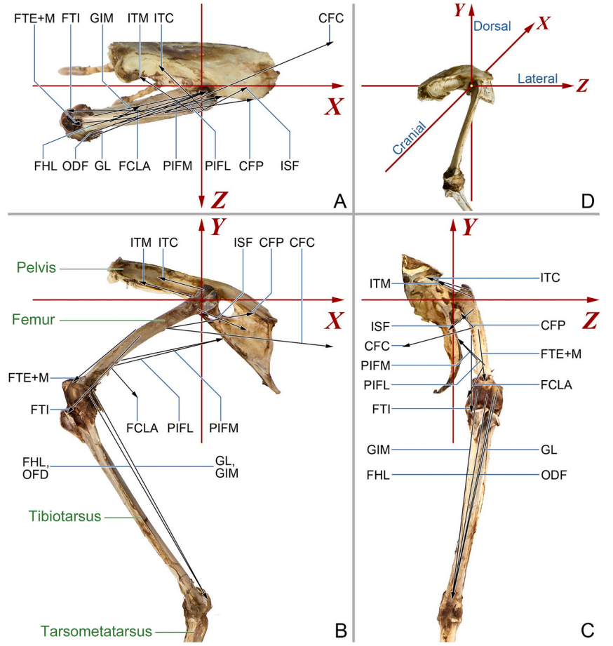
  • The hindlimbs play a crucial role in bird locomotion, making the biomechanical properties of the musculoskeletal system in these limbs a focal point for researchers studying avian behaviour. However, a comprehensive analysis of the mechanical performance within the long bones of hindlimbs during locomotion remains lacking. In the present study, the strain and deformation of the femur of Cabot’s Tragopans (Tragopan caboti) were estimated. We employed inverse simulation to calculate the force and moment of femoral muscles during mid-stance terrestrial locomotion and conducted finite element analysis to calculate femoral strain. Results showed that during mid-stance, the femur experiences combined deformation primarily characterized by torsion, bending, and compression. It emphasises the importance of considering the influence of varying loads on bone adaptation when investigating bone form-function relationships. Muscles were found to play a significant role in offsetting joint loads on the femur, subsequently reducing the deformation and overall strain on the bone. This reduction enhances femoral safety during locomotion, allowing birds to meet mechanical demands while maintaining a lightweight bone structure. Notably, the M. iliotrochantericus caudalis significantly reduces torsional deformation of the proximal femur, protecting the vulnerable femoral neck from high fracture risk induced by rotation load. Given that the femur torsion during terrestrial locomotion in birds is associated with changes in hindlimb posture due to their adaptation to flight, the characteristics of M. iliotrochantericus caudalis may provide insight into the locomotor evolution of theropods and the origin of avian flight.

  • Among the four species of ground jays (Podoces) in the world, two are found in the west of China (Qian et al., 1965; Cheng, 1987): the Xinjiang Ground Jay (P. biddulphi) (Fig. 1) and the Mongolian Ground Jay (P. hendersoni). Xinjiang Ground Jays occur only in the Taklimakan Desert, the southern part of Xinjiang. Since the species was established by A. Hume in 1874, little has been known of its status and ecology. The current essay describes such information based on a long-term field survey.

    Figure  1.  The Xinjiang Ground Jay in the Lopnur Desert (Photo by Ming Ma)

    The ground jays are residents at the center of the Taklimakan Desert (37–42°N, 77–94°E, 790–1500 m elevations; Fig. 2). Most of distribution range of the species falls within Xinjiang, with a few extending to the east, e.g. the Qaidam Basin in Qinghai Province and Dunhuang in Gansu Province (Collar et al., 2001; Sun and Li, 2009). Interestingly, Mongolian Ground Jays are distributed around the range of Xinjiang Ground Jays (Fig. 2). Such a pattern should be a result of inter-species competition. There is evidence showing that the ground jays are expanding their range from west to east during recent decades (Ma, 2010).

    Figure  2.  Distribution of the Xinjiang Ground Jay and the Mongolian Ground Jay in the Taklimakan Desert

    Ground jays are well adapted to desert and semi-desert regions (Ludlow and Kinnear, 1933). Compared with crows or choughs, ground jays are sandy in plumage, presumably providing protection from desert predators (Londei, 2004). As their names indicate, ground jays spend much of their time on the ground. The strong legs should be adaptive to the habit. However, ground jays nest in shrubs and trees, a characteristic similar to that followed by the Corvids species.

    Information on breeding ecology of Xinjiang Ground Jays is very limited. A total of 20 ground jay nests were recorded in Niya and Qarqan from 2003 to 2004. The birds placed their nests on the small desert-poplar tree Populus diversifolia and Tamarix spp. bushes, averaging 1.09 ± 0.15 m (range = 0–2.30 m, n = 18) above the ground (Fig. 3). The nests were composed of sheep wool, camel's wool, horse's hair, dead leaves, dry grass, and the soft cottony growth of reeds, with poplar skin, twigs and small sticks being lined at the base. The external diameter of the nest is 35.75 ± 2.30 cm (range = 16–55 cm, n = 16), the internal diameter of the nest is 12.82 ± 0.85 cm (range = 9–20 cm, n = 14), and depth of the nest is 9.50 ± 1.00 cm (range = 5–16 cm, n = 13) and the height is 20.88 ± 1.33 cm (range = 12–35 cm, n = 16) (Fig. 4a). Clutch size varied between 1 and 3 eggs (1.89 ± 0.31 eggs, n = 9), the diameters of the eggs are 32.88 ± 0.83 mm × 23.48 ± 0.09 mm, and the weight of egg is 8.33 ± 0.88 g (n = 4). The color of egg is pale green and grayish white with brown spots scattered all over the surface, rather more densely at the broad end (Fig. 4b). The parents fed the young (Fig. 4c) more than 42 times during one day for one nest located in the middle March (Ma, 2004). After fledging, family flocks of 4 to 6 birds were encountered between early May and July. These data suggested that the ground jays laid eggs from late February to April.

    Figure  3.  Nest height above the ground in relation to nesting bush height of the Xinjiang Ground Jays
    Figure  4.  Breeding ecoloty of the Xinjiang Ground Jay. (a) The local guide in the field work, showing the nest built on a Tamarix bush; (b) The egg; (c) Feeding the chicks; (d) Nearly fledging chicks (photos by Ming Ma).

    Based on the transect counting conducted from 1988 to 2011, the density of ground jays was to be 3–5 pairs per 100 km2. It was estimated to have 4100–6700 pairs of ground jays over the species' range of 135000 km2.

    The ground jay populations seemed to drop during recent decades (Grimmett, 1991; Madge and Burn, 1994; Ma, 1998). However, with the intensified desertification in western China, the jays have a tendency to expand to the east.For example, there were some of new records in Gansu and Qinghai provinces (Collar et al., 2001; Sun and Li, 2009; Ma, 2010). Arguably, this bird is an indicator species of desertification and climate change.

    Human activities such as oil industry, ecotourism, land exploitation and overgrazing should be responsible for the population decline (Ma, 2001; Ma and Kwok, 2004). The restricted range and special requirements for nesting habitats suggest an urgent conservation need for this species. Now, although the bird is classified as "near-threatened" (Collar et al., 2001), no protection measures have been in practice (Zheng and Wang, 1998).

    The research is supported by the Science Supporting Project of National Ministry of Science and Technology (2008BAC39B04) and the National Natural Science Foundation of China (30270211, 30470262, 30970340). Sincere thanks are due to all participants in the field work, especially to Jinghe Gu, Kwok Hon Kai, Zexin Jia, Batuerhan, Eugene Potapov, Chuanbo Wang, Feng Xu, Yiqun Wu, Mike Kilburn, Geoff Carey, Richard Lewthwaite, Sebastien Lepetz and Paul Leader. Gratitudes are also given to Prof. Xin Lu from Wuhan University who helps revise the earlier manuscript very carefully.

  • Abe, S., Narra, N., Nikander, R., Hyttinen, J., Kouhia, R., Sievänen, H., 2016. Exercise loading history and femoral neck strength in a sideways fall: a three-dimensional finite element modeling study. Bone 92, 9–17.
    Abourachid, A., Höfling, E., 2012. The legs: a key to bird evolutionary success. J. Ornithol. 153, 193–198.
    Allen, V.R., Kilbourne, B.M., Hutchinson, J.R., 2021. The evolution of pelvic limb muscle moment arms in bird-line archosaurs. Sci. Adv. 7, eabe2778.
    Anderson, F.C., Pandy, M.G., 2001. Static and dynamic optimization solutions for gait are practically equivalent. J. Biomech. 34, 153–161.
    Bell, A., Chiappe, L.M., 2011. Statistical approach for inferring ecology of Mesozoic birds. J. Syst. Paleontol. 9, 119–133.
    Biewener, A.A., Dial, K.P., 1995. In vivo strain in the humerus of pigeons (Columba livia) during flight. J. Morphol. 225, 61–75.
    Biewener, A.A., Swartz, S.M., Bertram, J.E.A., 1986. Bone modeling during growth: dynamic strain equilibrium in the chick tibiotarsus. Calcif. Tissue Int. 39, 390–395.
    Bishop, P.J., Cuff, A.R., Hutchinson, J.R., 2021. How to build a dinosaur: musculoskeletal modeling and simulation of locomotor biomechanics in extinct animals. Paleobiology 47, 1–38.
    Bishop, P.J., Hocknull, S.A., Clemente, C.J., Hutchinson, J.R., Barrett, R.S., Lloyd, D.G., 2018. Cancellous bone and theropod dinosaur locomotion. Part Ⅱ-A new approach to inferring posture and locomotor biomechanics in extinct tetrapod vertebrates. PeerJ 6, e5779.
    Bright, J.A., 2014. A review of paleontological finite element models and their validity. J. Paleontol. 88, 760–769.
    Carrano, M.T., 1998. Locomotion in non-avian dinosaurs: integrating data from hindlimb kinematics, in vivo strains, and bone morphology. Paleobiology 24, 450–469.
    Carrano, M.T., 2000. Homoplasy and the evolution of dinosaur locomotion. Paleobiology 26, 489–512.
    Carrano, M.T., Biewener, A.A., 1999. Experimental alteration of limb posture in the chicken (Gallus gallus) and its bearing on the use of birds as analogs for dinosaur locomotion. J. Morphol. 240, 237–249.
    Carrier, D.R., Leon, L.R., 1990. Skeletal growth and function in the California gull (Larus californicus). J. Zool. 222, 375–389.
    Close, R.I., 1972. Dynamic properties of mammalian skeletal muscles. Physiol. Rev. 52, 129–197.
    Crawford, R.P., Cann, C.E., Keaveny, T.M., 2003. Finite element models predict in vitro vertebral body compressive strength better than quantitative computed tomography. Bone 33, 744–750.
    Cubo, J., Casinos, A., 1998. Biomechanical significance of cross-sectional geometry of avian long bones. Eur. J. Morphol. 36, 19–28.
    Cuff, A.R., Daley, M.A., Michel, K.B., Allen, V.R., Lamas, L.P., Adami, C., et al., 2019. Relating neuromuscular control to functional anatomy of limb muscles in extant archosaurs. J. Morphol. 280, 666–680.
    Currey, J.D., 2002. Bones: Structure and Mechanics. Princeton University Press, Princeton.
    Demes, B., 2007. In vivo bone strain and bone functional adaptation. Am. J. Phys. Anthropol. 133, 717–722.
    Demes, B., Qin, Y.-X., Stern Jr., J.T., Larson, S.G., Rubin, C.T., 2001. Patterns of strain in the macaque tibia during functional activity. Am. J. Phys. Anthropol. 116, 257–265.
    Duda, G.N., Heller, M., Albinger, J., Schulz, O., Schneider, E., Claes, L., 1998. Influence of muscle forces on femoral strain distribution. J. Biomech. 31, 841–846.
    Duda, G.N., Schneider, E., Chao, E.Y.S., 1997. Internal forces and moments in the femur during walking. J. Biomech. 30, 933–941.
    Erdemir, A., McLean, S., Herzog, W., van den Bogert, A.J., 2007. Model-based estimation of muscle forces exerted during movements. Clin. Biomech. 22, 131–154.
    Falk, A.R., Lamsdell, J.C., Gong, E., 2021. Principal component analysis of avian hind limb and foot morphometrics and the relationship between ecology and phylogeny. Paleobiology 47, 314–336.
    Farke, A.A., Alicea, J., 2009. Femoral strength and posture in terrestrial birds and non-avian theropods. Anat. Rec. 292, 1406–1411.
    Ford, C.M., Keaveny, T.M., Hayes, W.C., 1996. The effect of impact direction on the structural capacity of the proximal femur during falls. J. Bone Miner. Res. 11, 377–383.
    Gatesy, S.M., 1990. Caudofemoral musculature and the evolution of theropod locomotion. Paleobiology 16, 170–186.
    Gatesy, S.M., 1999. Guineafowl hind limb function. Ⅱ: electromyographic analysis and motor pattern evolution. J. Morphol. 240, 127–142.
    Gatesy, S.M., Middleton, K.M., 1997. Bipedalism, flight, and the evolution of theropod locomotor diversity. J. Vertebr. Paleontol. 17, 308–329.
    Geier, A., Kebbach, M., Soodmand, E., Woernle, C., Kluess, D., Bader, R., 2019. Neuro-musculoskeletal flexible multibody simulation yields a framework for efficient bone failure risk assessment. Sci. Rep. 9, 6928.
    Goetz, J.E., Derrick, T.R., Pedersen, D.R., Robinson, D.A., Conzemius, M.G., Baer, T.E., et al., 2008. Hip joint contact force in the emu (Dromaius novaehollandiae) during normal level walking. J. Biomech. 41, 770–778.
    Goodno, B.J., Gere, J.M., 2018. Mechanics of Materials, ninth ed. Cengage Learning, Boston.
    Habib, M.B., Ruff, C., 2008. The effects of locomotion on the structural characteristics of avian limb bones. Zool. J. Linn. Soc. 153, 601–624.
    Hutchinson, J.R., Allen, V., 2009. The evolutionary continuum of limb function from early theropods to birds. Naturwissenschaften 96, 423–448.
    Hutchinson, J.R., Gatesy, S.M., 2000. Adductors, abductors, and the evolution of archosaur locomotion. Paleobiology 26, 734–751.
    Hutchinson, J.R., Rankin, J.W., Rubenson, J., Rosenbluth, K.H., Siston, R.A., Delp, S.L., 2015. Musculoskeletal modelling of an ostrich (Struthio camelus) pelvic limb: influence of limb orientation on muscular capacity during locomotion. PeerJ 3, e1001.
    Jepsen, K.J., Silva, M.J., Vashishth, D., Guo, X.E., van der Meulen, M.C., 2015. Establishing biomechanical mechanisms in mouse models: practical guidelines for systematically evaluating phenotypic changes in the diaphyses of long bones. J. Bone Miner. Res. 30, 951–966.
    Judex, S., Carlson, K.J., 2009. Is bone’s response to mechanical signals dominated by gravitational loading? Med. Sci. Sports Exerc. 41, 2037–2043.
    Khassetarash, A., Haider, I., Baggaley, M., Edwards, W.B., 2022. Tibial strains during prolonged downhill running: a finite element analysis. J. Biomech. Eng. 145, 041007.
    Krahl, A., Lipphaus, A., Sander, M.P., Maffucci, F., Hochscheid, S., Witzel, U., 2020. Humerus osteology, myology, and finite element structure analysis of Cheloniidae. Anat. Rec. 303, 2177–2191.
    Lee, J., Sim, H., Jeong, J., Kim, S.-Y., Yang, S., Jeong, S., et al., 2020. Biomechanical analysis of canine medial patellar luxation with femoral varus deformity using a computer model. BMC Vet. Res. 16, 471.
    Lieberman, D.E., Polk, J.D., Demes, B., 2004. Predicting long bone loading from cross-sectional geometry. Am. J. Phys. Anthropol. 123, 156–171.
    Lipphaus, A., Witzel, U., 2019. Biomechanical study of the development of long bones: finite element structure synthesis of the human second proximal phalanx under growth conditions. Anat. Rec. 302, 1389–1398.
    Lutz, F., Mastel, R., Runge, M., Stief, F., Schmidt, A., Meurer, A., et al., 2016. Calculation of muscle forces during normal gait under consideration of femoral bending moments. Med. Eng. Phys. 38, 1008–1015.
    Main, R.P., Biewener, A.A., 2004. Ontogenetic patterns of limb loading, in vivo bone strains and growth in the goat radius. J. Exp. Biol. 207, 2577–2588.
    Main, R.P., Biewener, A.A., 2006. In vivo bone strain and ontogenetic growth patterns in relation to life-history strategies and performance in two vertebrate taxa: goats and emu. Physiol. Biochem. Zool. 79, 57–72.
    Main, R.P., Biewener, A.A., 2007. Skeletal strain patterns and growth in the emu hindlimb during ontogeny. J. Exp. Biol. 210, 2676–2690.
    Marsh, R.L., Ellerby, D.J., Carr, J.A., Henry, H.T., Buchanan, C.I., 2004. Partitioning the energetics of walking and running: swinging the limbs is expensive. Science 303, 80–83.
    Martelli, S., Pivonka, P., Ebeling, P.R., 2014. Femoral shaft strains during daily activities: implications for atypical femoral fractures. Clin. Biomech. 29, 869–876.
    Meilak, E.A., Provini, P., Palmer, C., Gostling, N.J., Heller, M.O., 2021. Unpublished results. On the hindlimb biomechanics of the avian take-off leap. bioRxiv.
    Mendez, J., Keys, A., 1960. Density and composition of mammalian muscle. Metabolism 9, 184–188.
    Milne, N., O’Higgins, P., 2012. Scaling of form and function in the xenarthran femur: a 100-fold increase in body mass is mitigated by repositioning of the third trochanter. Proc. Biol. Sci. 279, 3449–3456.
    Pelker, R.R., Friedlaender, G.E., Markham, T.C., Panjabi, M.M., Moen, C.J., 1983. Effects of freezing and freeze-drying on the biomechanical properties of rat bone. J. Orthop. Res. 1, 405–411.
    Rankin, J.W., Rubenson, J., Hutchinson, J.R., 2016. Inferring muscle functional roles of the ostrich pelvic limb during walking and running using computer optimization. J. R. Soc. Interface 13, 20160035.
    Rayfield, E.J., 2007. Finite element analysis and understanding the biomechanics and evolution of living and fossil organisms. Annu. Rev. Earth Planet Sci. 35, 541–576.
    Reilly, S.M., Blob, R.W., 2003. Motor control of locomotor hindlimb posture in the American alligator (Alligator mississippiensis). J. Exp. Biol. 206, 4327–4340.
    Rudman, K.E., Aspden, R.M., Meakin, J.R., 2006. Compression or tension? The stress distribution in the proximal femur. Biomed. Eng. Online 5, 12.
    Shahar, R., Banks-Sills, L., 2002. Biomechanical analysis of the canine hind limb: calculation of forces during three-legged stance. Vet. J. 163, 240–250.
    Shahar, R., Banks-Sills, L., Eliasy, R., 2003. Stress and strain distribution in the intact canine femur: finite element analysis. Med. Eng. Phys. 25, 387–395.
    Smith, N.C., Payne, R.C., Jespers, K.J., Wilson, A.M., 2007. Muscle moment arms of pelvic limb muscles of the ostrich (Struthio camelus). J. Anat. 211, 313–324.
    Sverdlova, N.S., Witzel, U., 2010. Principles of determination and verification of muscle forces in the human musculoskeletal system: muscle forces to minimise bending stress. J. Biomech. 43, 387–396.
    Taylor, M.E., Tanner, K.E., Freeman, M.A.R., Yettram, A.L., 1996. Stress and strain distribution within the intact femur: compression or bending? Med. Eng. Phys. 18, 122–131.
    Ulivieri, F.M., Rinaudo, L., 2021. Beyond bone mineral density: a new dual X-ray absorptiometry index of bone strength to predict fragility fractures, the bone strain index. Front. Med. 7, 590139.
    Wang, L., Wei, X., Liang, X., Zhang, Z., 2021. Ontogenetic changes of hindlimb muscle mass in Cabot’s tragopan (Galliformes, Phasianidae) and their functional implications. Anat. Rec. 304, 2841–2855.
    Wei, X., Zhang, Z., 2019. Ontogenetic changes of geometrical and mechanical characteristics of the avian femur: a comparison between precocial and altricial birds. J. Anat. 235, 903–911.
    Wei, X., Zhang, Z., 2021. Femoral mechanical performance of precocial and altricial birds: a simulation study. Avian Res 12, 18.
    Witzel, U., Preuschoft, H., 2005. Finite-element model construction for the virtual synthesis of the skulls in vertebrates: case study of Diplodocus. Anat. Rec. 283, 391–401.
    Young, V.K.H., Wienands, C.E., Wilburn, B.P., Blob, R.W., 2017. Humeral loads during swimming and walking in turtles: implications for morphological change during aquatic reinvasions. J. Exp. Biol. 220, 3873–3877.
    Zeffer, A., Johansson, L.C., Marmebro, A., 2003. Functional correlation between habitat use and leg morphology in birds (Aves). Biol. J. Linn. Soc. 79, 461–484.
    Zhang, Z., Zheng, G., 1996. A study on the appendicular myology of the Cabot’s tragopan. In: Society, C.O., et al. (Ed.), Studies of Chinese Ornithology. China Forestry Publishing House, Beijing, pp. 210–216. (in Chinese with English abstract).
  • Related Articles

Catalog

    Figures(5)  /  Tables(1)

    Article Metrics

    Article views (9) PDF downloads (6) Cited by()

    /

    DownLoad:  Full-Size Img  PowerPoint
    Return
    Return